Lasermikroskope sind wichtige Werkzeuge in der Biologie
Die heutigen konfokalen Mikroskope, Multiphotonenmikroskope, Super-Resolution-Techniken wie PALM und STORM, Lichtblatttechniken und vieles mehr sind alle auf Laser angewiesen, um die Grundlagen des Lebens auf zellulärer Ebene zu entschlüsseln.
19. Oktober 2021 von Coherent
Die Verwendung des Lichtmikroskops in den Biowissenschaften geht auf den niederländischen Geschäftsmann Anton van Leeuwenhoek zurück, der damit berühmt wurde, weil er damit die ersten Einblicke in winzige Lebensformen erhielt. Spulen Sie etwa 400 Jahre zurück, und Wissenschaftler in allen Bereichen der Biowissenschaften verlassen sich immer noch auf optische Mikroskope als wichtige Werkzeuge für ihre Arbeit. Das ist in jeder Hinsicht eine ziemlich beeindruckende Langlebigkeit. Und das Wort „mikroskopisch“ ist natürlich seit Langem ein allgemein akzeptierter Begriff für alles, was klein ist.
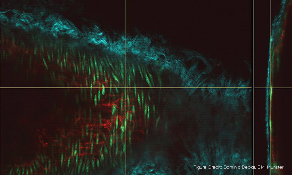
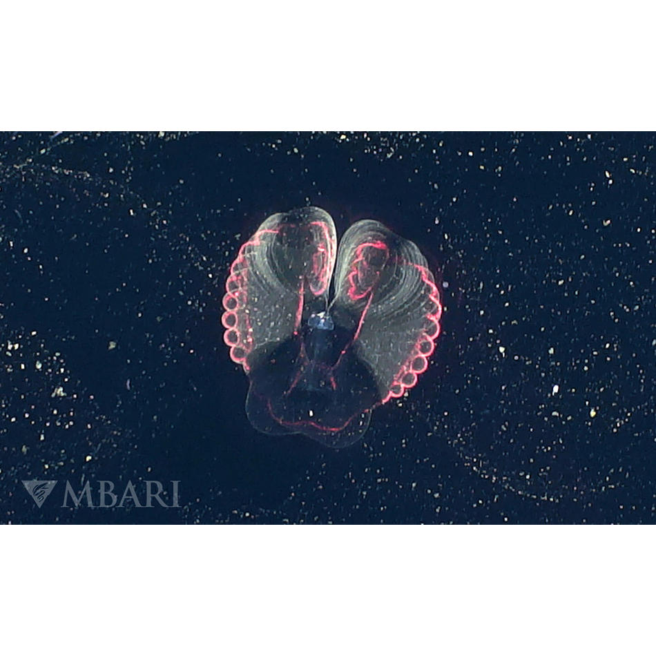
3D-Mikroskopie in Aktion. Eine Halsschlagader der Maus, in der mehrere Zellkomponenten mit zwei Wellenlängen eines Chameleon Discovery Lasers als unterschiedliche Farben dargestellt werden. Das Hauptbild zeigt die Halsschlagader „en face“ und der vertikale Schnitt auf der rechten Seite ist eine orthogonale Ansicht der Arterienwand.
Das Leben mit Mikroskopen kartieren
Warum ist das Lichtmikroskop immer noch so wichtig und was hat das mit Lasern zu tun? Nun, es ist das einfachste Instrument, um die Struktur von kleinen Dingen abzubilden. Aber genauso wichtig ist, dass es sich zu dem einzigen Instrument entwickelt hat, das Ihnen sagen kann, woraus diese Strukturen bestehen. Durch die Manipulation und Messung der Farben (Wellenlängen) des Lichts, das mit einer Probe interagiert, können Wissenschaftler eine Karte aller Arten von verschiedenen Biochemikalien erstellen. Und da jeder lebende Organismus, von einer einzelligen Amöbe über einen Baum bis hin zu einem Elefanten, auf komplexen biochemischen Reaktionen beruht, ist das eine ziemlich nützliche Fähigkeit.
Dieses chemische Mapping war früher recht grob. Vor hundert Jahren wurde eine tote („fixierte“) Probe einer Pflanze oder eines Tieres chemisch mit einem Farbstoff behandelt, den man Beize nannte. Dies könnte sich an das gesamte Fett in der Probe heften, oder vielleicht auch an das gesamte Protein. Der Betrachter konnte dann sehen, welche Teile der Probe durch diesen Farbstoff gefärbt waren und welche nicht.
Die Wissenschaftler von heute haben unzählige Farbstoffe zur Auswahl und ihre Studien sind viel, viel anspruchsvoller. Die meisten von ihnen sind fluoreszierende Chemikalien, die oft als Fluorophore oder Fluorochrome bezeichnet werden. (Fluoreszierende Materialien absorbieren Licht bei einer Wellenlänge und geben das Licht bei einer anderen, längeren Wellenlänge wieder ab.) Manche Fluorophore sind einfach nur Chemikalien, die aus der Flasche kommen, aber oft handelt es sich um fluoreszierende Proteine, die die Pflanze oder das Tier gentechnisch verändert hat, um sie direkt selbst zu produzieren.
Ein Laser ist die ultimative Lichtquelle für Mikroskope
Aber was ist mit Lasern? Nun, dazu kommen wir jetzt. Es hat sich herausgestellt, dass der Laser aus mehreren Gründen die ultimative Lichtquelle für die Fluoreszenzmikroskopie ist. Wissenschaftler wollen Mikroskope einsetzen, um immer mehr Details zu sehen. Wir nennen das räumliche Auflösung, oder einfach nur Auflösung. Sie wollen auch echte 3-dimensionale Dinge sehen, nicht nur dünne, tote Scheiben. Und sie wollen auch Mikroskope verwenden, um biologische Vorgänge in Echtzeit zu beobachten. Laser haben sich als die Lichtquelle erwiesen, die all das möglich macht.
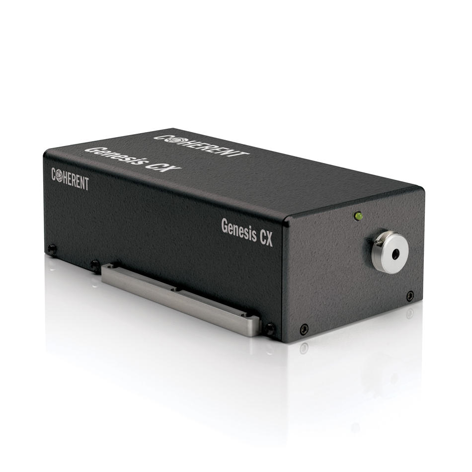
Laser bringen der Fluoreszenzmikroskopie mehrere entscheidende Vorteile. Erstens emittiert ein Laser Licht mit nur einer Wellenlänge. Und auch dank unserer optisch gepumpten Halbleitertechnologie (OPSL) kann diese Wellenlänge so gewählt werden, dass sie der Absorption bestimmter Fluorophore entspricht. Glasfilter vor der Kamera des Mikroskops blockieren dann das von der Probe gestreute Laserlicht (d. h. die Blendung) und sorgen dafür, dass nur die Fluoreszenz selektiv abgebildet wird. Jetzt können Sie ein einzelnes Wellenlängenband mit Hilfe einer Lampe oder einer LED mit einem Filter erhalten. Aber der Laserstrahl kann auf einen viel kleineren Punkt fokussiert werden als das Licht einer Lampe oder LED. Dies ist der Schlüssel zum konfokalen Laser-Scanning-Mikroskop (CLSM), das 3D-Bilder ohne die unscharfe Hintergrundfluoreszenz liefert, die dies sonst unmöglich machen würde.
Laser können auch eine viel höhere Intensität liefern als eine Lampe oder LED, so dass selbst schwache Fluoreszenz schnell abgebildet werden kann. Außerdem ist die hohe und einstellbare Intensität des Lasers der Schlüssel zu den neuesten Super-Resolution-Techniken (z. B. PALM, STORM), die Bilder mit einer Auflösung von bis zu einigen Nanometern erzeugen. Und das ist eine große Sache, die 2014 mit dem Nobelpreis ausgezeichnet wurde, denn noch vor 40 Jahren dachte man, dass man mit einem sichtbaren Mikroskop niemals über die harte Beugungsgrenze, d. h. etwa 250 Nanometer, hinausgehen könnte.
Live dabei sein
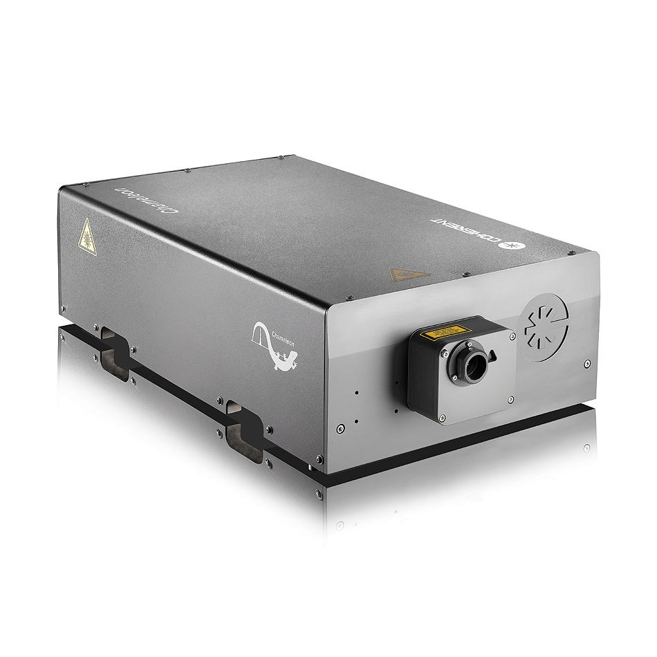
In vielen Bereichen der Biowissenschaften, vor allem in den Neurowissenschaften, ist den Forschern die Bildtiefe ebenso wichtig wie die Auflösung. Sie wollen scharfe Bilder tief im Gewebe oder sogar im Inneren ganzer Organismen erhalten. Und sie wollen dies mit lebendem Gewebe tun. Während konfokale und superauflösende Methoden für fixierte Proben hervorragend geeignet sind, verursachen sie bei lebenden Proben in der Regel zu viel Photodestruktion. Glücklicherweise gibt es eine Technik namens Multiphotonenmikroskopie, die diese Herausforderung mit Hilfe von ultrafast Lasern meistert. (Noch ein Nobelpreis!). Tatsächlich ist dies eine so wichtige Anwendung für ultrafast Laser, dass Coherent eine ganze Familie von Chameleon Ultrafast Lasern herstellt, die speziell auf die Multiphotonenmikroskopie zugeschnitten sind.
Mit Blick auf die Zukunft entwickeln Wissenschaftler auf der Grundlage der Multiphotonenmikroskopie Werkzeuge für medizinische Anwendungen wie Echtzeit-Biopsien. Das ist möglich, denn einige der Multiphotonen-Methoden benötigen nicht einmal einen Farbstoff oder ein fluoreszierendes Protein! Dies wird als markierungsfreie Bildgebung bezeichnet.
Dies ist also nur ein mikroskopischer Einblick in die enormen Auswirkungen des Lasermikroskops in der Biologie. Wir würden gerne glauben, dass van Leeuwenhoek sehr erstaunt und beeindruckt wäre, denn das sind wir auch!
Verwandte Ressourcen