CUSTOMER SUCCESS STORY
Chameleon Flexibility and Reliability Key to Lab Productivity
The Challenge
The photonic imaging facility (IMAG’IC) is part of Institut Cochin (Inserm U1016, CNRS UMR 8104, and Université Paris Cité UMR-S1016), which is a large biomedical research center in Paris, France. IMAG’IC is well-equipped for virtually every type of optical microscopy with a total of 15 different microscopes. It is an open-access collaborative facility enabling researchers from within Institut Cochin and elsewhere to perform cutting-edge studies using state-of-the-art equipment. This includes research on diverse samples like cells or tissues, both in vitro and in vivo. Many of the instruments are located in biosafety levels 2 and 3 laboratories, which allows research and imaging of live cells, tissue samples of human origin and/or infected with pathogens, and whole mammals infected with communicable diseases.
Dr. Thomas Guilbert is a Research Engineer within IMAG’IC who explains, “We’ve had a multiphoton microscope for about 15 years including a tunable (Chameleon Ultra II) ultrafast laser, mainly used for SHG experiments. We recently obtained funding to acquire a second multiphoton microscope to expand the range of our imaging solutions and needed to select the optimum laser for this. In particular, we wanted a laser that would support the widest range of non-linear imaging techniques, including short and long-wavelength fluorescent probes, as well as label-free techniques like endogenous fluorescence, harmonic generation (SHG, THG) imaging, and even Fluorescence Lifetime Imaging (FLIM) methods. We also wanted a laser that would be easy to operate and highly reliable, in order to support our dense schedule of users (~250 per year) at IMAG’IC with varying levels of skill and experience in advanced microscopy methods.”
The Solution
The IMAG’IC team considered several lasers for the new microscope and eventually chose a Coherent Chameleon Discovery. Thomas states that there were several factors that made this laser ideal for their needs. He notes that “The broad spectral bandwidth – 660 nm to 1320 nm – means that we can perfectly match the excitation wavelength requirements for all common probes and transgenic proteins as well as for label-free techniques. In a core facility, you must adapt quickly to different types of experiments, from harmonic imaging on thin unlabeled slides to intravital microscopy of small mammals”. He also cites having a second synchronous beam at 1040 nm as highly useful for experiments needing simultaneous excitation at two wavelengths as well as for stimulated Raman.
Thomas adds that, “We really need exceptional reliability to support an intense program of collaborative research and we knew from our long experience with the Ultra II that Chameleon lasers deliver that, and even with the Mira 900 that I used a lot during my PhD work. We also wanted the wide range of pre-compensation that comes with Discovery. Plus we needed short pulse width and high peak power to maximize imaging depth in thick tissues and even to perform some ablation.” He explains that some researchers are using the laser to ablate inside samples of collagen gel to make synthetic model blood vessels for example. And lastly, he adds that having a laser with pulses synchronized with external detectors enables techniques such as FLIM imaging and FRET-FLIM, or to easily separate two fluorophores having the same spectral emission.
The Result
Thomas states that the flexibility and reliability of Discovery means that the new microscope has been widely used, with over 6 papers resulting from just its first 20 months of operation, and more to come. These include microscopy of organ development, various skin diseases and care methods, microscopy of cleared, ex-vivo, and live tissues including lung, kidney, and brain. A particularly novel set of studies entail imaging the migration of immune cells in live slices of tumors. Thomas himself is often involved in the research workflow, such as for the fibrosis studies led by Profs Yannick Allanore and Frédéric Batteux whose research is focused on pathogenesis and innovative therapies in chronic fibro-inflammatory diseases such as systemic sclerosis, rheumatoid arthritis, and endometriosis. SHG imaging is ideal for this type of translational research, because it selectively images collagen, the major component involved in fibrosis, and of course it requires no labels.
“We really need exceptional reliability to support an intense program of collaborative research and our Chameleon lasers deliver that."
— Thomas Guilbert, Research Engineer, Institut Cochin, IMAG’IC, Paris, France
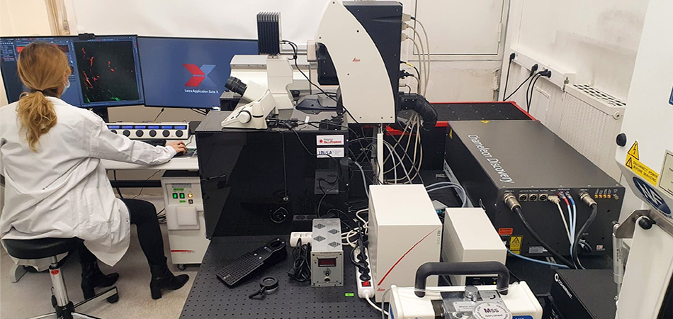
Chameleon Discovery dual laser coupled to a Leica Microsystems Dive microscope with NDD spectral detection and Falcon module in a biosafety level 2 lab. IMAG’IC core facility, Institut Cochin, Paris, France.
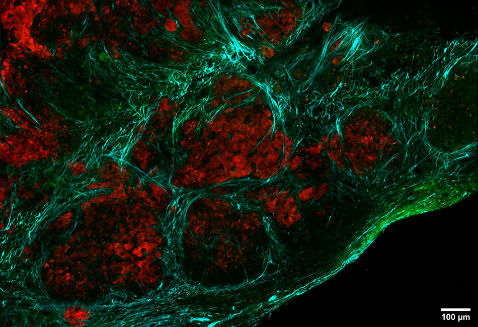
Multiphotonic imaging of thick slice of tumor tissue. SHG (cyan), TPEF (green), and mCherry tumor cells (red). Dr Emmanuel Donnadieu and IMAG’IC collaboration.